
FSPR vs SDR: What's the Difference? A Detailed Comparison

Two Names, One Goal: Understanding the Difference
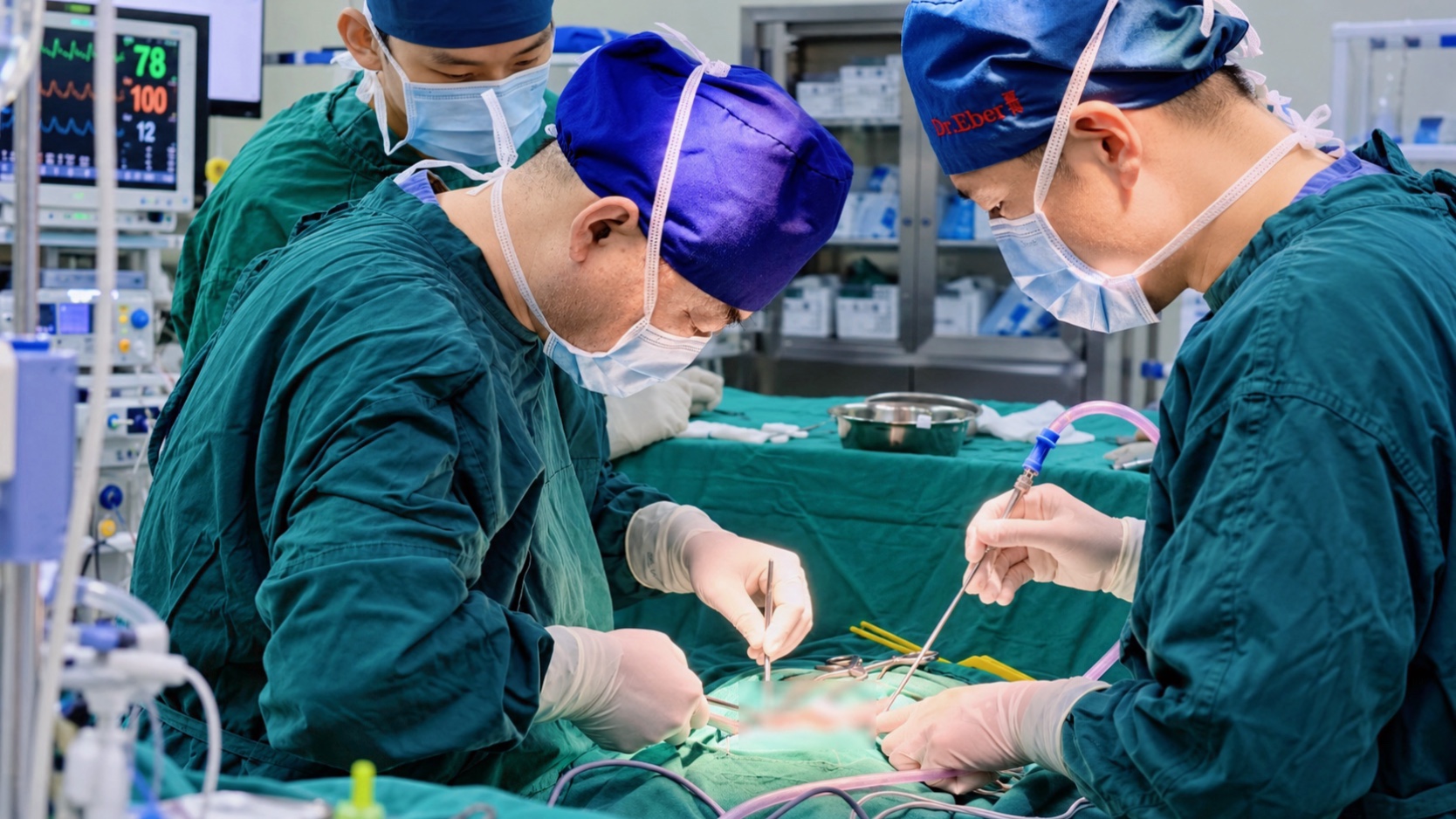
If you have researched cerebral palsy surgery, you have likely encountered both FSPR and SDR. These terms are often used interchangeably, but they represent important differences in surgical technique and precision. Understanding the distinction is critical for making an informed decision about your child's treatment. At Eber Neurosurgery, we perform FSPR — the more advanced and precise version of the procedure — pioneered and refined by Dr. Sun Chengyan over 20+ years and 30,000+ procedures.
Table of Contents
Origins and History

1912: Otfrid Foerster
German neurophysiologist first described rhizotomy for spasticity, cutting sensory nerve roots to relieve muscle tone.
1970s-80s: SDR Developed
Dr. Thomas Peacock and Dr. T.S. Park refine SDR (Selective Dorsal Rhizotomy), cutting portions of sensory nerve roots.
1990s: SDR Standardized
SDR becomes standard in US/UK for spastic cerebral palsy. Single-level or multi-level approaches used without intraoperative electrophysiological monitoring.
2002+: FSPR in China
Dr. Sun Chengyan at Huashan Hospital develops Functional Selective Posterior Rhizotomy, adding multi-level intraoperative electrophysiological monitoring.
2007: Gold Standard
FSPR becomes the 'gold standard' for cerebral palsy surgical indications, widely adopted in China.
2015-Present: FSPR Refined
Dr. Sun's team performs 400+ FSPR surgeries annually, with continuous refinement. FSPR becomes the standard in China.
Surgical Technique: The Key Difference

Pre-treatment
Post-treatment
The fundamental difference between SDR and FSPR lies in how nerve fibers are selected for cutting. Traditional SDR relies primarily on anatomical identification — the surgeon visually identifies nerve rootlets and cuts a predetermined percentage (typically 50-70%) based on appearance and tactile feedback. FSPR adds multi-level intraoperative electrophysiological monitoring: each nerve rootlet is electrically stimulated, and the EMG response is measured to identify precisely which fibers are causing abnormal spasticity. Only those functionally abnormal fibers are selectively cut — typically 30-50% of fibers — preserving more normal nerve function.
Detailed Comparison
| Feature | Traditional SDR | FSPR (Eber Technique) |
|---|---|---|
| Full Name | Selective Dorsal Rhizotomy | Functional Selective Posterior Rhizotomy |
| Nerve Selection | Anatomical (visual) | Electrophysiological (EMG-guided) |
| Fibers Cut | 50-70% (predetermined) | 30-50% (functionally abnormal only) |
| Monitoring | Minimal or none | Multi-level intraoperative EMG |
| Precision | Moderate — may cut normal fibers | High — only abnormal fibers cut |
| Normal Nerve Risk | Higher (up to 50-70%) | Lower (<30%) |
| Spasticity Reduction | Good (60-80%) | Excellent (80-95%) |
| Recovery | Similar | Similar or slightly faster |
| Surgeon Skill | Requires SDR training | Requires specialized monitoring equipment |
Why Electrophysiological Monitoring Matters
The addition of electrophysiological monitoring is not a minor refinement — it fundamentally changes the precision of the procedure. In traditional SDR, the surgeon estimates which fibers to cut based on experience. In FSPR, each nerve rootlet is stimulated with electrical impulses, and the resulting muscle response is measured via EMG. Fibers that produce abnormal, widespread, or sustained responses are identified as pathological and selectively cut. Fibers that produce normal, localized responses are preserved. This means: less normal nerve tissue is sacrificed, lower risk of sensory loss, more precise spasticity reduction, and better preservation of bladder and bowel function.
Traditional SDR
- Experience-based
- May sacrifice normal fibers
- Less precise fiber selection
- Higher sensory loss risk
- May cut normal fibers
- Limited to surgeon's experience
FSPR (Eber)
- EMG-guided precision
- Only abnormal fibers cut
- Lower complication risk
- Preserves sensory function
- Preserves bladder & bowel
- Requires specialized monitoring equipment
Outcomes: What the Data Shows
Both SDR and FSPR produce significant and lasting spasticity reduction. Studies of SDR show 60-80% spasticity reduction at 5-year follow-up (long-term). Eber's FSPR data shows 80-95% spasticity reduction with lower complication rates. The most important predictor of outcome is not SDR vs FSPR, but surgeon experience and post-operative rehabilitation intensity. A high-volume surgeon with integrated rehabilitation produces better outcomes regardless of technique name.
“The difference between SDR and FSPR is the difference between estimating and measuring. With electrophysiological monitoring, we do not guess which fibers cause spasticity — we see it on the EMG. This precision is why our complication rates are so low.”
Frequently Asked Questions
A:Yes. We encourage pre-treatment visits when possible. We also offer virtual tours.
A:Shanghai Eber Hospital is a Class 2A designated institution (China's highest).
A:International menus are available on request. Halal, kosher, and other dietary needs are accommodated.
A:Yes. Companion beds are available in patient rooms, and we can arrange nearby apartments.
Have Questions? Talk to Your Dedicated International Coordinator
Free consultation, no obligation. We respond within 48 hours.
Ready to Start Your Recovery Journey?
Submit your case — Dr. Sun Chengyan's team responds within 48 hours
Medical Review by Sun Chengyan, MD, PhD — Chief Neurosurgeon, Shanghai Eber Hospital
Last Updated: September 2026 | Content reviewed for medical accuracy